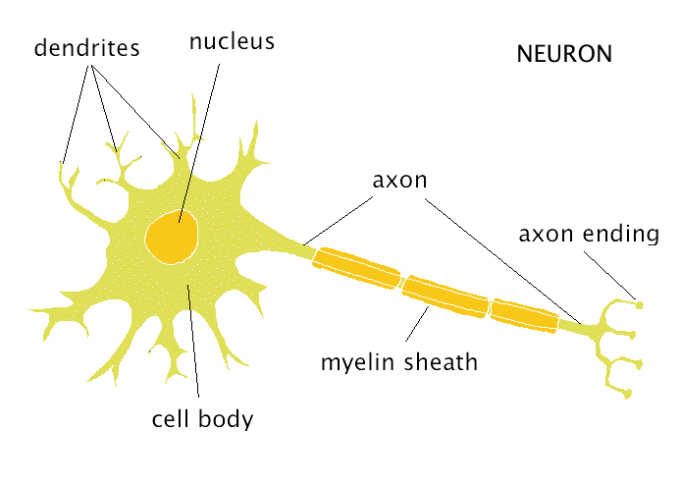
Each layer within the respective region of the brain contains specific neurons. The former has six layers, while the latter has three layers. The cerebrum (neocortex) and cerebellum are histologically divided into layers. These cells are associated with proprioception and joint position. This process bifurcates close to the cell body and one branch (the central, or axonic) travels from the cell body to the spinal cord, while the other (the peripheral or dendritic) travels from the periphery to the cell body. The cell body, which is found in dorsal root ganglia, has one single process that serves both the role of the axon and the role of the dendrite. Pseudounipolar cells relate to an older nomenclature used to describe unipolar cells. These cells are usually encountered in peripheral nerves and sensory ganglia. They have a single axon projecting from the spherical cell body, while other regions of the cell membrane are devoid of dendritic branches. Unipolar cells are another subtype of neurons. These cells are found in the vestibulocochlear (hearing), olfactory and ocular (viz. They have a single axon projecting from one end of the oval cell body and a lone dendritic tree extending from the other end. Bipolarīipolar cells are only associated with afferent impulses. Because of the numerous branching, the cells appear fusiform or polygonal. These cell types have a single axon extending from one end of the cell body and several dendrites branching as they protrude from the other side of the cell body. Multipolar cells are most predominant in the brain and spinal cord and are inclusive of motor neurons as well as interneurons. The cells can either be multipolar, bipolar, unipolar or pseudounipolar. Neurons can also be classified based on the number of processes that emerge from the somata. The activity of the receptors can be modified by other neurotransmitters like glutamate (excitatory response) or glycine and GABA (inhibitory response).

Cholinergic receptors that are stimulated by acetylcholine (ACh) include muscarinic (M) and nicotinic (N) receptors.Catecholaminergic receptors such as α-, β-receptors and dopamine (D) receptors are activated by norepinephrine and dopamine, respectively.There are numerous receptors that respond to the varying neurotransmitters that are found in the synaptic clefts: Neurotransmitters can be classified as amino acids (glycine, gamma-aminobutyric acid and glutamate), catecholamines (norepinephrine or noradrenaline and dopamine), or cholines (acetylcholine). It will also cover briefly the histological layers of the central and peripheral nervous systems.

This article will explain the histology of neurons, providing you with information about their structure, types, and clinical relevance. Peripheral nerves - epineurium, perineurium, endoneurium Myelinated (nodes of Ranvier) vs nonmyelinatedĬerebrum - molecular, external granular, external pyramidal, internal granular, internal pyramidal, multiform layersĬerebellum - molecular, Purkinje, granular layers Presynaptic terminal, synaptic cleft, neurotransmitter, postsynaptic terminalĬell body (soma), dendrites (covered with dendritic spines), axon, axon hillock. 'bottom-up' information, in the opposite direction carrying sensory information the central nervous system, via afferent neurons (e.g. to alert the body of danger).
to permit locomotion) (efferent neurons) or, 'top-down' information from the brain to the periphery, via efferent neurons (e.g.The cytology of a neuron facilitates the transmission of either: This laid the foundation for what has been referred to as the neuron doctrine.Ī neuron (nerve cell) is a specialized cell that conveys electrochemical impulses throughout the body. Subsequent research showed that each neuron is capable of operating independently. Prior to the late 19th century, neurons were viewed as collective functional units that formed a syncytium.


 0 kommentar(er)
